Ear Treatment

EMBRYOLOGY OF EAR

Embryology is the study of the origin and development of single individual embryo in prenatal period.

Development of Different Parts of the Ear

1) Outer Ear Development -
Image
  • By the end of 4th week, the auricle develops from 6 mesenchymal proliferations (hillocks) of 1st and 2nd pharyngeal arches, surrounding the first pharyngeal cleft.
  • These auricular hillocks, three on each side of the external meatus, later fuse and form the definitive auricle.
  • External auditory meatus develops from ectoderm of first pharyngeal cleft.
  • At the 3rd month, epithelial cells at the bottom of the meatus proliferate, forming a solid epithelial plate, the meatal plug.
  • In the 7th month, this plug dissolves and the epithelial lining of the floor of the meatus participates in formation of the definitive eardrum.
  • TM has three layers. Inner layer “endoderm”, Middle layer “mesoderm” and Outer layer “ectoderm”.
2) Middle Ear Development

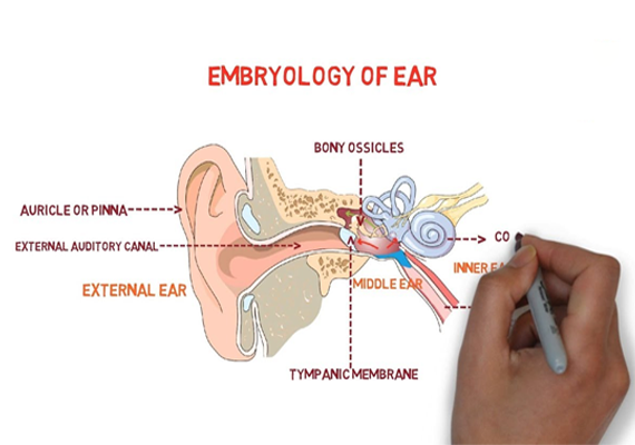
The middle ear cavity and the auditory tube arise from first pharyngeal pouch called the tubotympanic sulcus, lined by endoderm.

  • By the end of 7th week, the mesenchyme above the primitive tympanic cavity shows a number of condensations caused by proliferation of the dorsal tips of pharyngeal arches I and II.
  • The condensations become the cartilaginous precursors of the auditory ossicles, the malleus, the incus, and the stapes.
  • The malleus and incus are derived from pharyngeal arch I (Meckel’s cartilage) and the stapes is derived from arch II (Reichert’s cartilage).
  • The ossicles appear during the first half of fetal life but remain embedded in mesenchyme until 8th month.
  • After birth, epithelium of the tympanic cavity invades bone of the developing mastoid process. Later, most of the mastoid air sacs come in contact with the antrum and tympanic cavity.
  • In the 7th month, this plug dissolves and the epithelial lining of the floor of the meatus participates in formation of the definitive eardrum.
Image
3) Inner Ear and the Formation of the Membranous Labyrinth
  • Membranous labyrinth: Derived from ectodermal invagination from otic placode.
  • Bony labyrinth: Arises from mesoderm.
  • In the 7th month, this plug dissolves and the epithelial lining of the floor of the meatus participates in formation of the definitive eardrum.

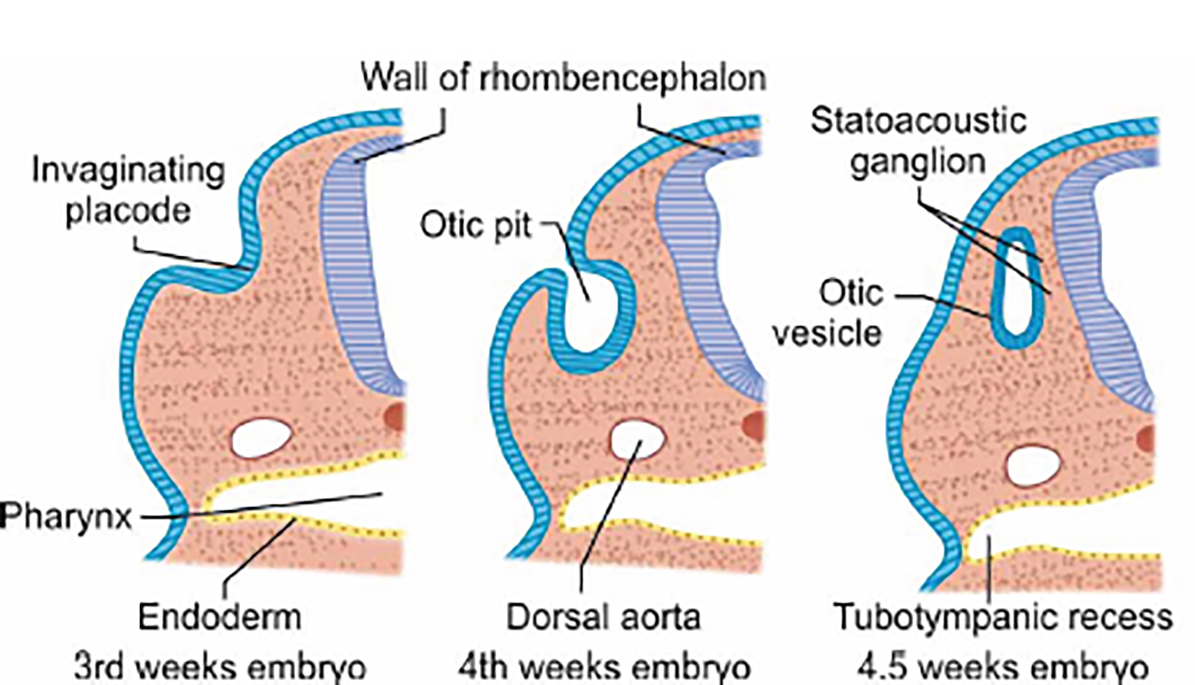
Early in the 4th week, a thickening of the surface ectoderm on each side of the hindbrain called otic placode forms otic pit and finally form the otic (auditory) vesicle. Membranous labyrinth arises from this ectodermal invagination from otic placode.

Book Your Appoinment