
Cystic swelling arising in connection with persistent portion of a part or whole of ectoderm lined cervical sinus.
Along the anterior border of sternomastoid usually in the region of the angle of jaw. There is a slight left-sided predominance.
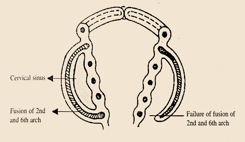
The cause is usually a development abnormality arising in the early prenatal period, typically failure of obliteration of the second, third, and fourth branchial cleft, i.e. failure of fusion of the second branchial arches and epicardial ridge in lower part of the neck. The appendages of the ectodermal lined enclosed space (cervical sinus), sweat and sebaceous glands continue their secretions which gradually distends the space to form the cyst. If sinus tracts are present, they typically drain into the tonsillar fossa, found between the palatoglossal arch and the palatopharyngeal arch.

Tooth paste like material rich in cholesterol.
The cyst wall is composed of squamous epithelium (90%), columnar cells with or without cilia, or a mixture of both with lymphoid infiltrate, often with prominent germinal centers and few subcapsular lymph sinuses. Cholesterol crystals may be found in the fluid extracted from a branchial cyst.
Superficial to the Internal carotid, glossopharyngeal nerve, stylopharyngeus, muscle. (Structures derived from the third arch). Deep to the lesser cornu of the hyoid bone, stylohyoid ligament, posterior belly of digastric muscle, facial nerve and external carotid artery. (Structures derived from the third arch).