Ear Treatment

CONDUCTIVE HEARING LOSS

Image

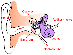
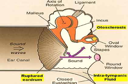
This type of hearing loss can occur due to any condition which interferes with the conduction of sound reaching the cochlea. The lesion may lie in the external ear, tympanic membrane, middle ear or ossicles up to stapediovestibular joint (oval window).

Aetiology:

The conductive hearing loss may be congenital or acquired.

  • Congenital
  • Congenital cholesteatoma.
  • Stapes fixation.
  • Malleus head fixation.
Acquired

    External Ear:

  • Wax.
  • Foreign body.
  • Furuncle.
  • Acute inflammation.
  • Exostosis of ear canal.

    Middle Ear:

  • Tympanic membrane perforation.
  • Conditions having fluid in the middle ear, Hemotympanum, Acute otitis media, Secretory otitis media.
  • Ossicular fixation.
  • Eustachian tube blockage.
  • Tumors of middle ear.
Image
Clinical Assessment of Hearing Loss:

Tuning fork tests

  • Rinne test:
    Bone conduction is better than air conduction in conductive hearing loss. It is also called Rinne negative. It is done with 256 Hz, 512 Hz and 1024 Hz tuning forks. 512 Hz tuning fork is most commonly used as it falls in the mid-speech frequency and the minimum overtones and undertones.
  • Weber test:
    It is done with 512 Hz tuning fork. The sound lateralizes to the worse affected ear in pure conductive hearing loss.
  • Absolute bone conduction test:
    This test is done to differentiate between conductive and sensorineural hearing loss. It is normal in conductive hearing loss.

Pure Tone Audiometry:

  • It is a qualitative and quantitative analysis of patients hearing.
  • It is helpful for medico legal purposes.
  • Helps in evaluating the pre and post-operative result.
  • Useful in malingering

Impedance Audiometry:

Useful in intact tympanic membrane to know the cause of conductive hearing loss.
1. Impedance audiometry: Useful in intact tympanic membrane to know the cause of conductive hearing loss.
2. Management: Most cases of conductive hearing loss can be managed by medical or surgical means.