Ear Treatment

MACEWEN’S TRIANGLE

Introduction:

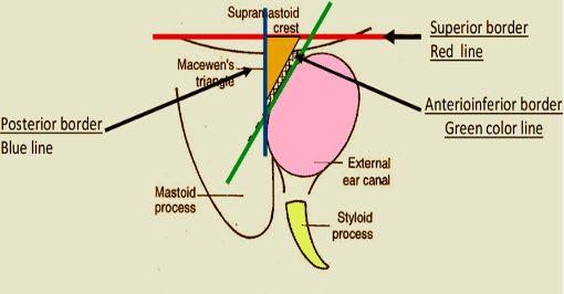
This triangle is the most important surgical landmark for the mastoid antrum or the largest mastoid air cell. In the temporal bone it is situated between the posterior wall of the external acoustic meatus and the posterior root of the zygomatic process. It is also called

  • Suprameatal triangle.
  • Suprameatal pit.
  • Mastoid fossa.
  • Foveola suprameatica.
History:

It is named after a Scottish surgeon, Sir William MacEwen (1864-1924).

Boundaries of this triangle:
  • Superiorly– Suprameatal crest.
  • Antero-inferiorly– posterosuperior segment of bony external auditory canal.
  • Posteriorly– A tangential line drawn from the posterior canal wall cutting the suprameatal crest.
Image
Related Pathology:
  • The mastoid antrum lies 1.5 to 2 cm deep to the triangle in the adult.
  • MacEwen’s Triangle is characterized by presence of multiple small perforating vessels; hence it is also known as cribrose (cribriform) area.
  • Henle’s spine which marks the anterior and inferior limit of dissection in a canal wall up mastoidectomy, is a content of MacEwen’s Triangle.
  • This is an important landmark while performing a cortical mastoidectomy.
  • The triangle lies deep to the cymba conchae.
  • The cortical bone is particularly thin at this area, making a preferred route for subperiosteal abscess (mastoid) formation, secondary to coalescent mastoiditis.