Otosclerosis
Otosclerosis is a hereditary localized disease originating from the otic capsule in which mature lamellar bone is removed by osteoclasts and replaced by woven bone of greater thickness, cellularity and vascularity. There is commonly a fixation in the stapediovestibular joint which fails or reduces the sound conduction resulting in deafness.
Aetiology :
- Age of onset:
Hearing loss usually starts between 20 and 30 years of age and is rare before 10 and after 40 years.
- Sex:
Females are affected twice as often as males but in India, otosclerosis seems to predominate in males.
- Race:
White races are affected more than black Americans. It is common in Indians but rare among Chinese and Japanese.
- Genetic Predisposition:
Mutations and altered expression of the SERPINF1 gene is seen in patients with familial otosclerosis. This gene encodes PEDF (pigment epithelium-derived factor) which is a known regulator of bone density and is the first disease associated gene to be identified in otosclerosis. Otosclerosis may be associated with osteogenesis imperfecta with history of multiple fractures. The triad of symptoms of osteogenesis imperfecta, otosclerosis and blue sclera is called Van der Hoeve syndrome. Lesions of otic capsule seen in osteogenesis imperfecta are histologically indistinguishable from those of otosclerosis and both are due to COL1A1 or COL1A2 genes encoding type I collagen.
- Viral infection:
Persistent viral infection of the otic capsule may cause otosclerosis. Otosclerosis is seen in Paget’s disease, which has viral aetiology. Increased expression of specific measles virus receptor CD46 isoforms is seen in otosclerotic footplates.
- Autoimmune Disease:
Otosclerosis represents a form of autoimmune disease with humoral autoimmunity to type II collagen.
- Cytokines:
Cytokines like transforming growth factor β1 (TGF-β1) and bone morphogenetic protein (BMP) are associated with otosclerosis.
- Hormonal factors:
Since angiotensin II stimulates the secretion of TNF-α, the renin–angiotensin-aldosterone system (RAAS) may play a role in the regulation of bone remodelling.
- Effect of other factors:
Hearing loss due to otosclerosis may be initiated or made worse by pregnancy. Similarly, deafness may increase during menopause, after an accident or a major operation.
Clinical Features :
- Deafness:
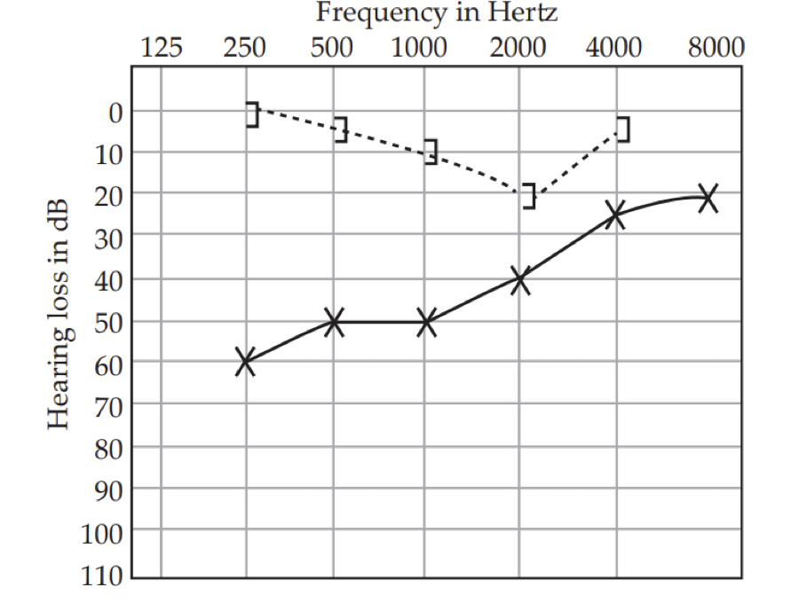
The deafness is usually bilateral occurring between the third and fifth decade. Audiometry shows a typical conductive hearing loss with dip in bone conduction curve at 2000 Hz called Carhart notch. An Impedance audiometry confirms the ossicular fixation.
- Tinnitus:
It is an indication of sensorineural hearing loss which is seen in patients having this disease since a long period.
- Vertigo:
These attacks are usually transient as a result of toxic enzymes.
- Occasionally, a reddish hue may be seen on the promontory through the tympanic membrane (Schwartz sign). This is indicative of active focus with increased vascularity.
Treatment
- Medical line of treatment: Oral sodium fluoride therapy can be given 50 mg daily for a period of 6 months to 1 year, depending on the tolerance level of the patient. However, this therapy is usually for medically unfit patients and results are unreliable.
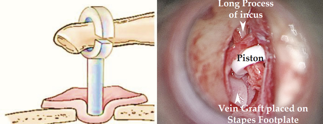
- Surgical treatment: Stapedotomy.