LARYNX

LARYNGOCELE

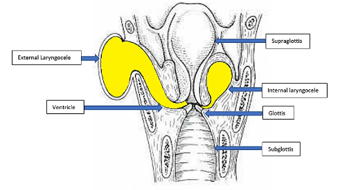
These are air filled cystic swellings due to dilatation of the saccule lined by columnar ciliated epithelium.

Types:

A laryngocele may be:
  1. Internal which is confined within the larynx and presents as distension of false cord and aryepiglottic fold. 
  2. External in which distended saccule herniates through the thyroid membrane and presents in neck.
  3. Combined or mixed in which both internal and external components are seen

Aetiology:
It is supposed to arise from raised transglottic air pressure
  1. Seen in professional trumpet players, glass blowers.
  2. Persons having chronic cough and asthma.
  3. A laryngocele in an adult may be associated with carcinoma which causes obstruction of saccule.
Clinical features:
  1. Swelling in the neck which reduces on pressure and increases on Valsalva manoeuvre.
  2. Hoarseness of voice with normal vocal cords.
  3. Sudden onset stridor.
  4. Dysphagia with halitosis.
  5. Snoring.
  • 6. Pain.
  • 7. Coughing.
  • 8. Boyce sign– Gurgling sound on compressing the external laryngocele with reduction of swelling.
  • 9. Pyocoele formation can be a sequelae.

Investigations:
  1. Plain X-ray neck shows an air-filled sac.
  2. CT scan neck shows the extent and origin of the laryngocele. Both the above investigations are done with and without Valsalva’s manoeuvre.   
Treatment:
  1. Surgical treatment is advised only if patient is symptomatic or if the laryngocele is infected.
  2. Surgery aims at excising the saccule.
  3. Tracheotomy is done as an emergency in cases of stridor.

Surgery for Internal laryngocele includes:
       
  1. Transthyrohyoid membrane approach.
  2.    
  3. V-shaped thyrotomy.
In Combined laryngocele:

External part is managed by
  • 1. Transthyrohyoid membrane approach.
  • 2. Thyrotomy with resection of the upper 1/3 thyroid cartilage.
  • 3. V-shaped thyrotomy.

Internal part is managed by
  1. Microlaryngoscopic CO2 laser resection.
  2. Endoscopic robotic surgery.
Book Your Appointment