ORAL CAVITY AND PHARYNX

LEUKOPLAKIA

White patch or plaque of squamous epithelium that cannot be characterized clinically or pathologically as any other disease.

Most common sites:
  • Buccal mucosa.
  • Oral commissure.
  • Ventral surface of tongue.
  • Floor of mouth.
  • Rarely, larynx.
Aetiology

1. Predisposing factors:

  • Sharp tooth.
  • Smoking.
  • Sepsis.
  • Spirits (Alcohol).
  • Spices.
  • Syphilis.
  • Ill-fitting dentures.
  • Plummer Vinson syndrome.
  • Oral submucous fibrosis.
  • Chronic hyperplastic candidiasis.

2. Age:
Can occur in any age but middle-aged (fourth decade) or elderly are more commonly affected.

3. Sex:
Males more frequently affected than females due to smoking and alcohol habits.

Types of leukoplakia:

Homogenous leukoplakia

Characterized by white patch – surface may be smooth or wrinkled.

Speckled or nodular leukoplakia

White patches or nodules or nodules on erythematous base.

Treatment of leukoplakia:
  1. Removal of predisposing cause.
  2. Oral antioxidants are useful.
  3. Excision Biopsy of the suspicious white patch.
  4. Small superficial patches of leucoplakia may be satisfactorily treated by CO2 laser excision, laser vaporization, photodynamic therapy and cryosurgery.
  5. Patients should be examined at regular three-month intervals to note appearance of warty excrescence. Any doubtful lesions if present must be removed and examined histologically.
  6. Radiotherapy: Not commonly given as it may increase the chances of malignancy.
Image

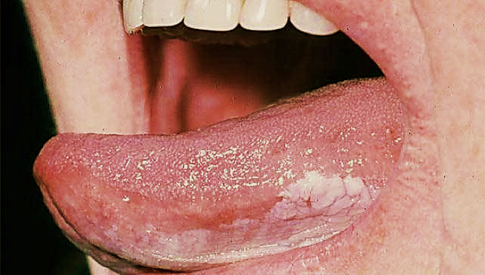
Leukoplakia at left lateral border of tongue.

Differential diagnosis:
  • Lichen planus.
  • Discoid lupus erythematosus.
  • White spongy nevus.
  • Candidiasis.
Book Your Appointment